Download Text as MSWord 6 document
Figure Legends
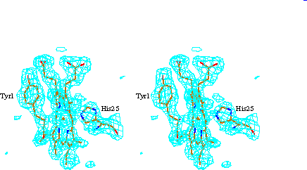
Figure 1. Omit map of the heme region. The electron density map is made with coefficients of Fo-Fc and F c, where Fc and F c are amplitudes and phases calculated from the final refined model after deleting the heme and residues Y1 and H25 to avoid model bias in the heme region. The map is contoured at the 3 s level.
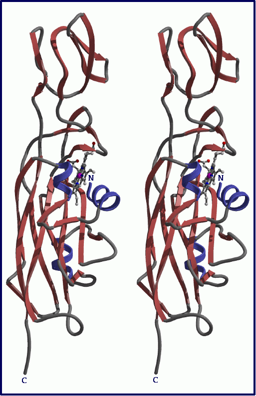
Figure 2. Stereo ribbon diagram of cytochrome f from C. reinhardtii (Raster 3D [Merrit, 1994 #104] rendered). In the wild type the C-terminus would be continuous forming a membrane spanning a -helix and a short cytoplasmic segment.
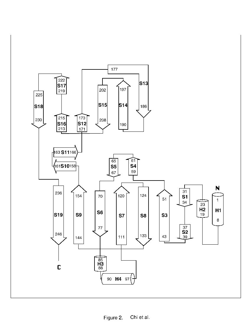
Figure 3. Schematic drawing of the secondary structure arrangement in the C. reinhardtii truncated cytochrome f.
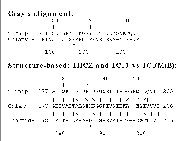
Figure 4. Sequence alignment of C. reinhardtii (Chlamy) with turnip cytochrome f based on Gray's alignment [Gray, 1992 #89], compared with alignment based on the structure (this work). In the latter, symbols between the lines indicate the closeness of superposition of the aligned residues, using the C. reinhardtii monomer B structure, turnip structure (entry 1CTM), and Phormidium laminosum structure (entry 1CI3) superimposed as described in the legend of Figure 5b: (|) Ca positions differ by less than 1.0 Å after alignment. (´ ) Ca positions differ by 1.0 Å or more. Insertions are placed to minimize the distance between Ca 's of aligned residues. The asterisk indicates the totally conserved carboxylate E186(turnip), E187(C. reinhardtii), and D187 [Gray, 1992 #89].
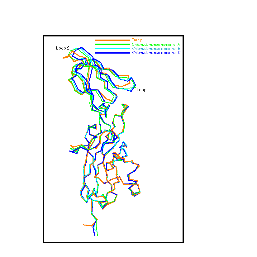
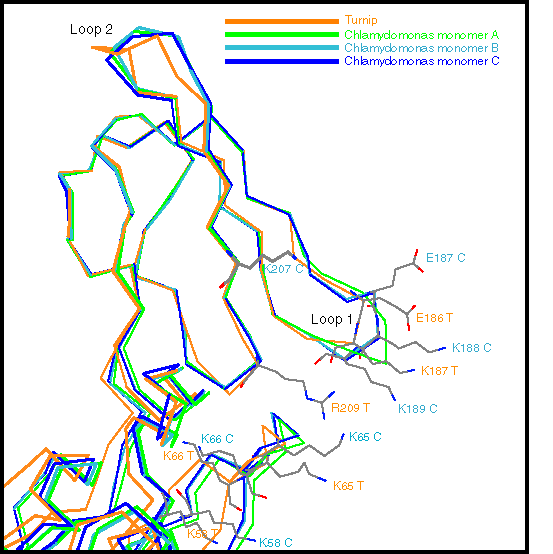
Figure 5. Comparison of the backbone representations of the three ncs-related monomers of C. reinhardtii (A, B, and C) and of turnip cytochrome f. The molecules were superimposed so as to minimize the rms distance between corresponding Ca . The "loop 1" and "loop 2" regions are discussed in the text.
a. The backbone of the full protein- Residues of the large domain 1-13, 18-169, and 231-248 of C. reinhardtii were matched with residues 1-13, 18-169, and 230-247 of turnip, respectively. For these 183 residues, the rms deviation in Ca position between the turnip cytochrome and the monomer B of C. reinhardtii cytochrome was 0.5 Å. The corresponding deviation of monomer A and monomer C with monomer B in C. reinhardtii were 0.3 Å, in both cases.
b. Detail of the small domain- Residues of the small domain 176-182, 191-197, and 204-222 of C. reinhardtii were matched with residues 176-182, 189-195, and 203-221 of turnip, respectively. For these 33 residues, the rms deviation in Ca position between the turnip cytochrome and the monomer B of C. reinhardtii cytochrome was 0.4 Å. The corresponding deviation of monomer A and monomer C with monomer B in C. reinhardtii were 0.2 Å, in both cases. Side chains for residues in the basic patch (putative plastocyanin binding site) are shown (with CPK coloring code) for the turnip structure and for monomer B of C. reinhardtii identified by T and C, respectively.
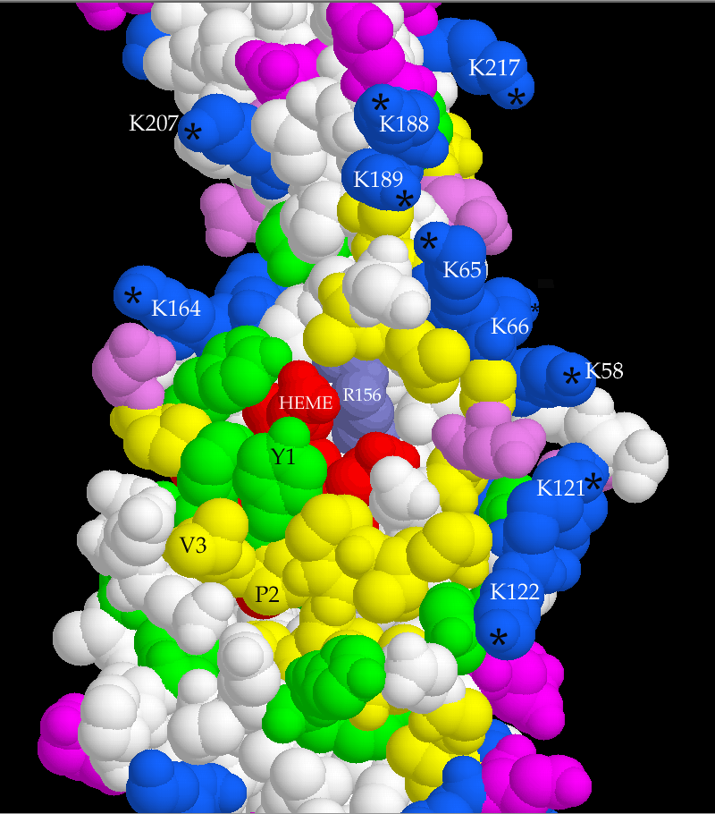
Figure 6. Space-filling Rasmol [Sayle, 1995 #106] representation of the central part of the C. reinhardtii truncated form of cytochrome f (monomer B). The hydrophobic outer face of the heme binding pocket (involving the N-terminus region) and the Lys cluster (putative plastocyanin binding site) are shown, see text. The top and base of the figure are areas distal and proximal to the membrane, respectively. Color code: blue, Lys; gray-blue, Arg; pink, Glu; magenta, Asp; yellow, 12 aliphatic, 4 Gly and 4 Pro residues; green, aromatic; white, polar; red, heme (only the two propionate chains are clearly seen in this view). Due to the molecule orientation, exposed water molecules W5-W7 are not seen. The Lys z N groups are identified with a star. Notice that K66 z N is not in the same plane with the other z N’s in the cluster and is almost not seen in this view. This figure encompasses approximately 2/3 of the large domain (bottom) and 1/2 of the small domain (top).
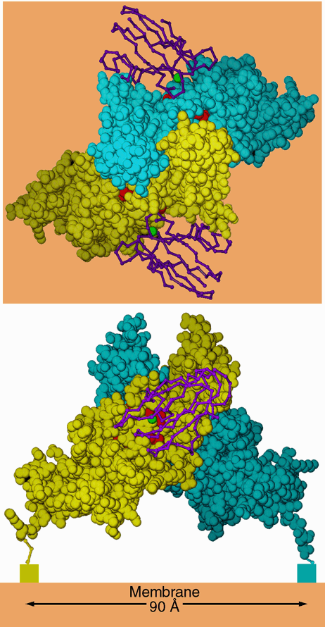
Figure 7. Dimer-like noncrystallographic symmetry observed in the crystal of C. reinhardtii cytochrome f. Above is a "top" view, looking down the 2-fold axis of symmetry. Below is a side view, looking parallel to a hypothetical membrane plane constructed perpendicular to the 2-fold (orange). The blue and yellow space-filling models are monomers A and B from the crystal of C. reinhardtii cytochrome f. The hemes are shown as red space-filling models. The rectangles below the C termini in the lower panel represent the lumenal ends of the transmembrane helices. The magenta backbone drawings are plastocyanin from the NMR structure of the plastocyanin-cytochrome f complex (2PCF), oriented with the operators that best superimpose cytochrome f of that structure on each monomer of the C. reinhardtii dimer. The copper atom of plastocyanin is shown as a green sphere. The interactions betwen these two monomers in the crystal may be similar to those involved in stabilizing a physiological dimer in vivo (see text). In this figure monomer B is taken directly from the structure 1CFM; monomer A is transformed by crystallographic symmetry operator [1/2+x, 1/2-y, 1-z] because the chosen asymmetric unit does not include this dimer.
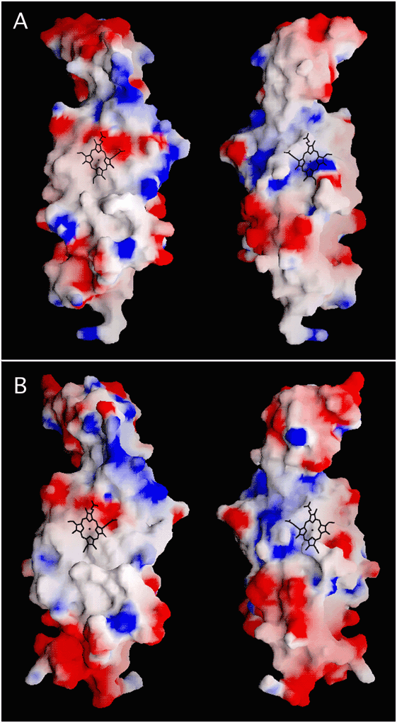
Figure 8. Electrostatic potentials at the molecular surface of the reduced forms of cytochromes f from C. reinhardtii (monomer B) (A) and from turnip (coordinates from 1CTM) (B). Two views rotated 180° around the longest dimension of the molecules are shown. The orientation of the molecules at the left panel is similar to the one shown in Figure 6. The surface electrostatic potentials are calculated by the program GRASP [Nicholls, 1991 #105] and displayed as a color gradient from red (electronegative, < -5 kT/charge) to blue (electropositive, >+5 kT/charge). The uncharged hydrophobic environment around the heme pocket and the overall positive potentials of the lysine cluster, with some differences between cytochromes f from two species, are seen. The dark stick figures indicate the location and orientation of the heme moiety, mainly buried in the protein (see Figure 6) with only the propionate groups exposed, if observed in the left view (see Figure 6), and one vinyl group exposed, if observed in the right view. In calculating electrostatic potentials, hydrogen atoms were omitted and their charge included in the charge of the atom to which they are bound (the default for GRASP). A charge of +1 was used for the Nz atoms of Lys, +0.5 for Nh 1 and Nh 2 of Arg, -0.5 for both carboxylate oxygens of Asp, Glu and the heme propionates. All other atoms of the heme were considered neutral, which is appropriate for the reduced heme.
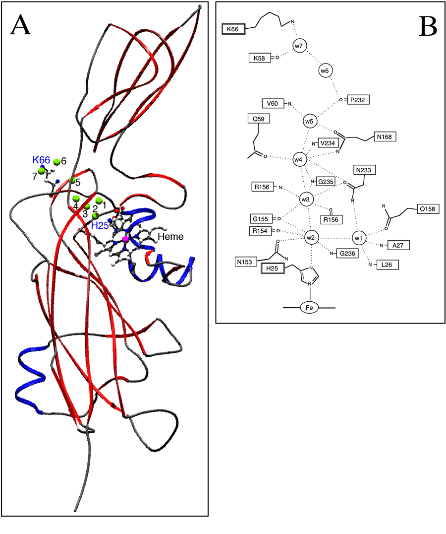
Figure 9. The "water chain" A) Locations of the seven water molecules forming the water chain (W1-W7, numbered green spheres; see text) are shown relative to the whole molecule. Residues H25 and K66, at the ends of the water chain, as well as the heme moieties are indicated in a ball-and-stick representation. B) Schematic representation of the hydrogen bonding network directly involving the buried (W1-W4) and exposed (W5-W7) water molecules of cytochrome f from C. reinhardtii. Side chains are not shown for those residues whose backbone atom is forming a hydrogen bond. All the residues shown in this network are strictly conserved in cytochromes f from C. reinhardtii and higher plants [Gray, 1992 #89].